Technology
Your workflow, streamlined.
Built by physicians to optimize ease-of-use — on serverless, HIPAA-compliant infrastructure purpose-built for medical imaging AI.
Prostate MRI AI
An automated biparametric MRI workflow: series selection, alignment, windowing, and prostate segmentation.
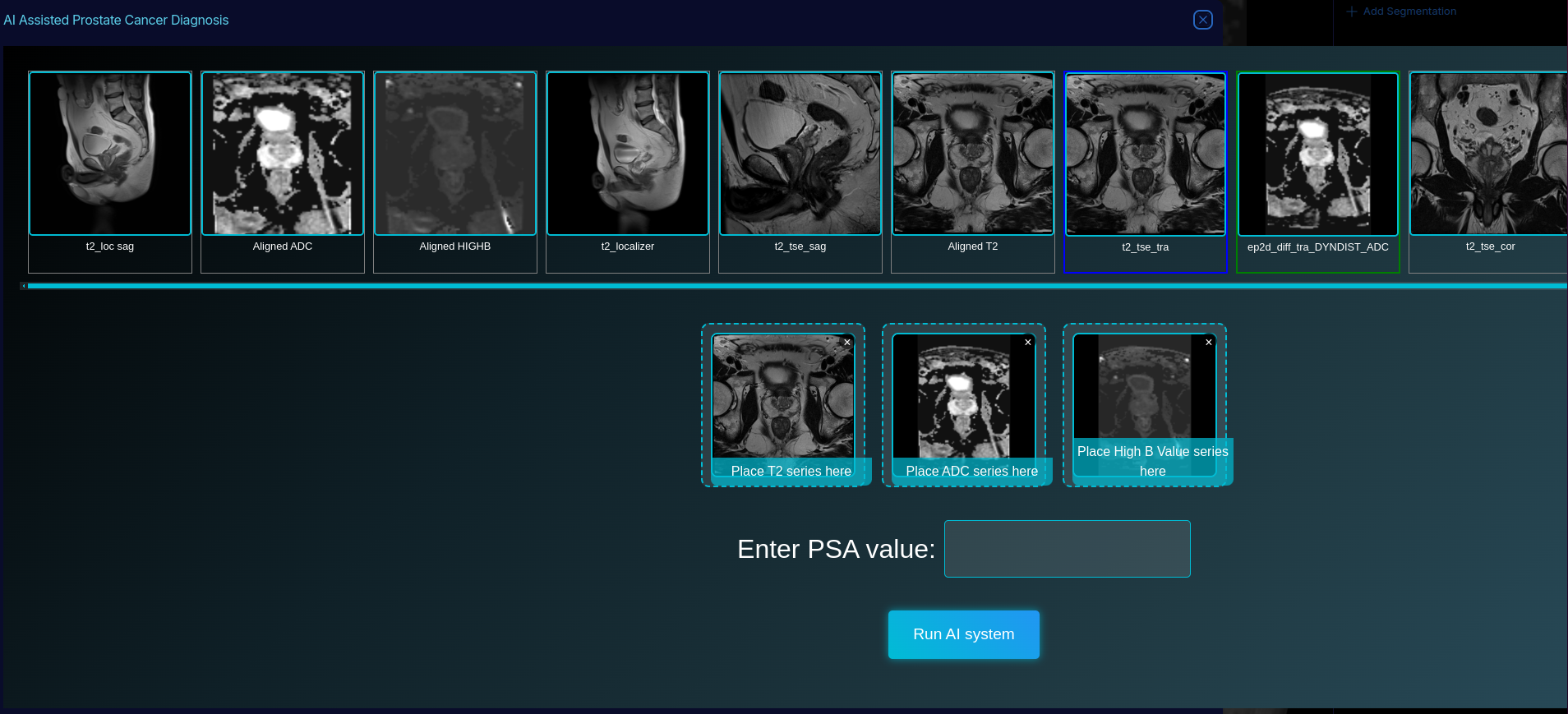
Automated Series Selection
Configure your institution's series naming conventions once. T2-weighted, ADC, and high b-value DWI series are then identified and populated automatically.
Automated Image Alignment
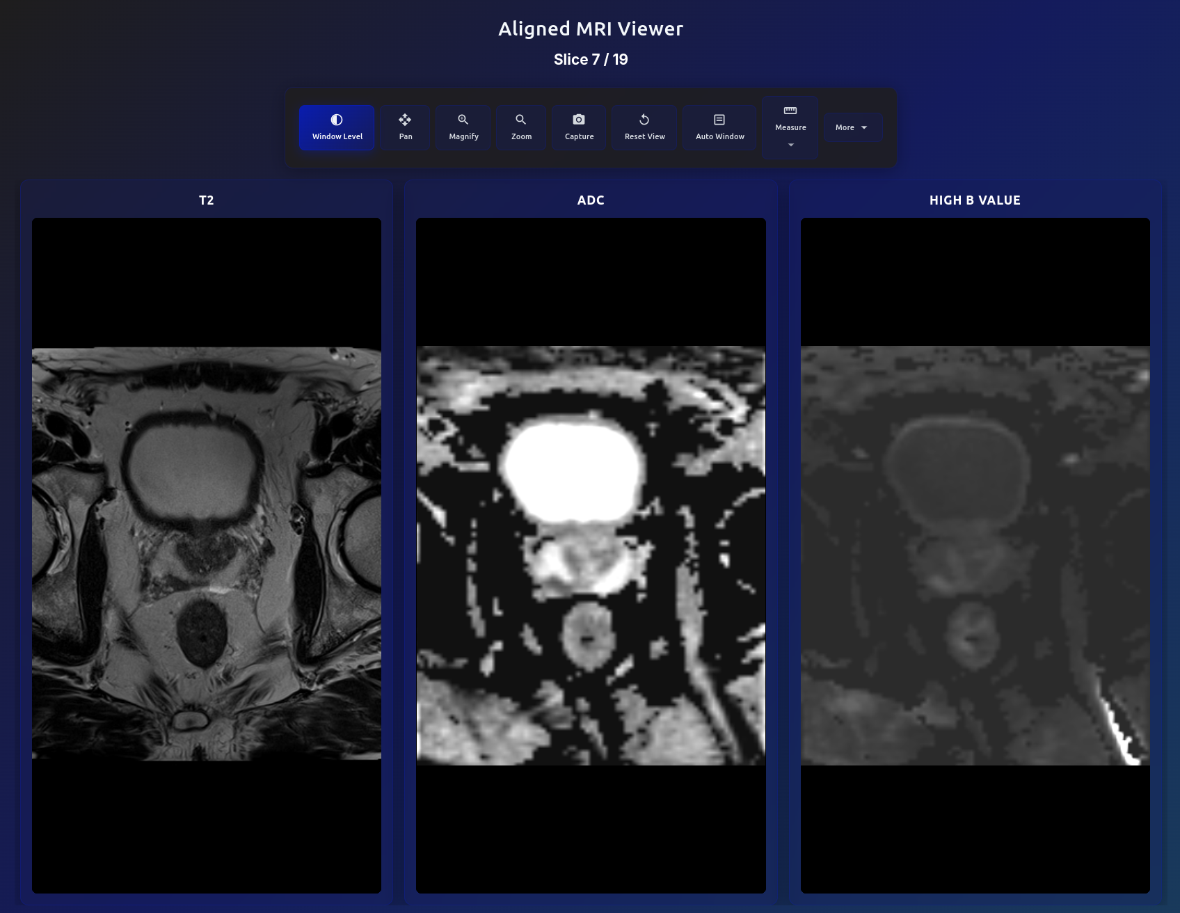
T2, ADC, and high b-value series are registered into the same voxel space. Scrolling stays synchronized across all three.
Automated Windowing
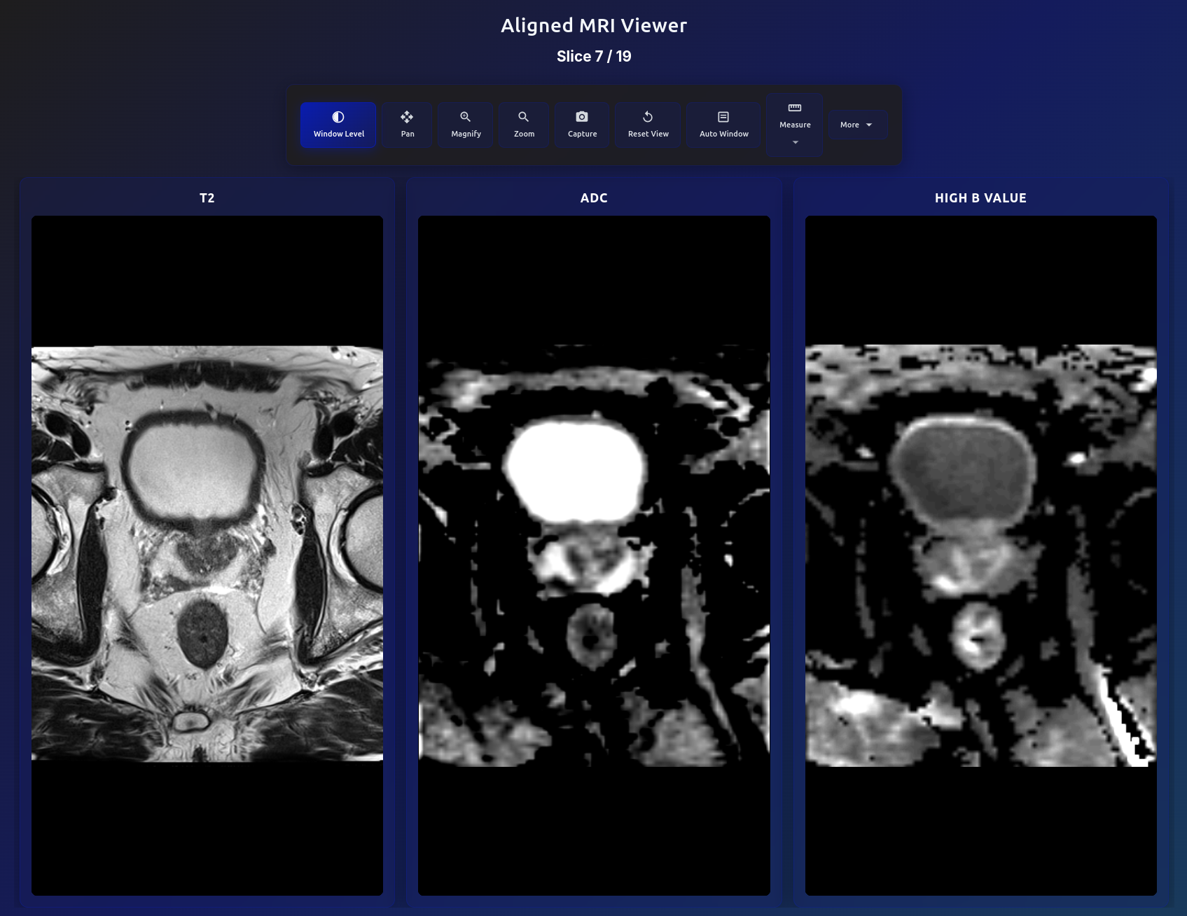
Window and level settings are applied automatically per sequence type, so studies are presented consistently without manual adjustment.
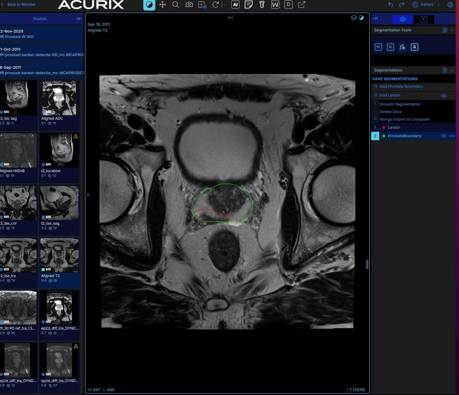
Prostate Segmentation
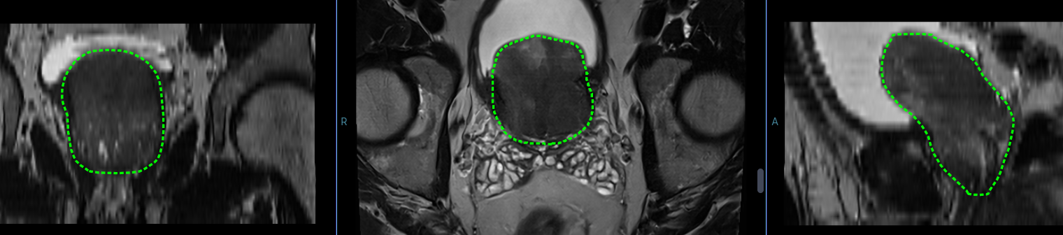
The prostate gland is segmented in three dimensions before the study is opened, using state-of-the-art neural networks running on cloud GPUs rather than local hardware — for the best chance of minimizing the need for user adjustment. Contours are delivered as DICOM RTSTRUCT objects and can be edited in the viewer or exported.
Lesion Annotation
Lesions are annotated by the reader, on the aligned T2, ADC, and high b-value images side by side. The alignment and auto-windowing help make the lesions identified by a radiologist obvious and the boundaries clear.
A target can be dropped as a sphere sized to the lesion, or traced freehand, and then refined in any plane. Targets are saved into the same DICOM RTSTRUCT as the gland segmentation, so a single object carries the whole study forward — to the report, to focal therapy planning, or to a fusion biopsy system.
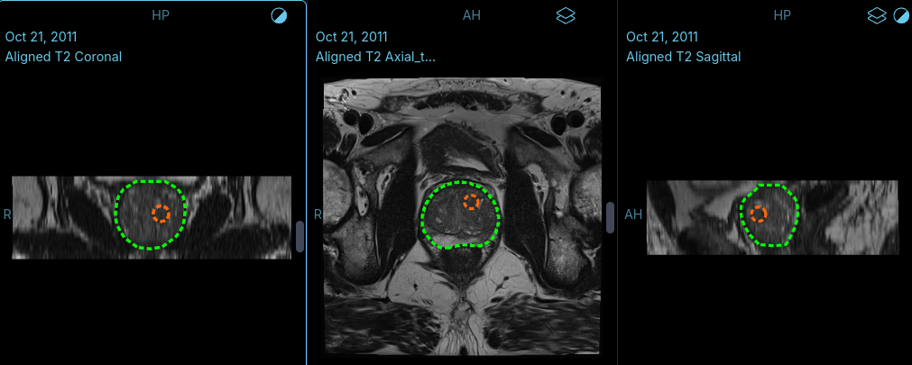
Multiplanar Adjustment
Contours can be edited in the axial, sagittal, or coronal view. The other two planes update from the edit, and edits can be undone.
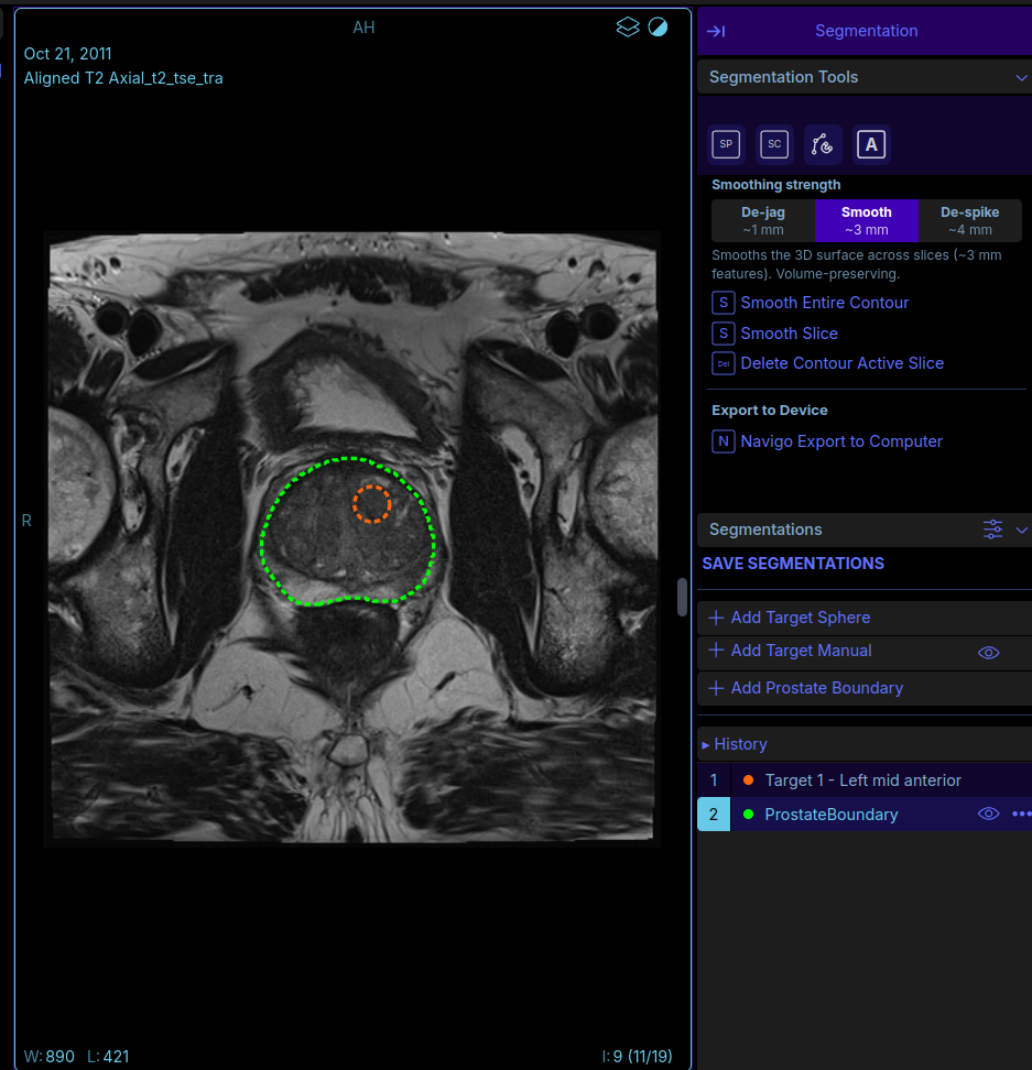
Intuitive Annotation Tools
Alignment, windowing, and segmentation are complete when the study opens, so the reader starts from a finished contour rather than a blank image.
Target placement and contour smoothing presets sit in one panel beside the image, and every edit can be undone.
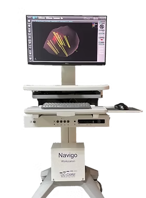
Navigo Fusion Biopsy Integration
Segmentation contours and lesion targets can be exported from the viewer to the UC-Care Navigo MRI/ultrasound fusion biopsy platform.
Upcoming
New AI capabilities currently in development on the Acurix platform.
PET/CT Viewer
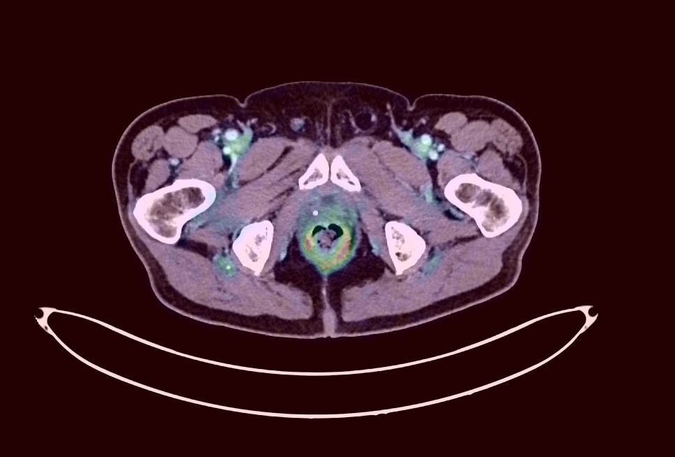
Native PET/CT viewing with true fusion display, SUV measurement, and Maximum Intensity Projection (MIP). Standard PACS systems often struggle to display PET scans natively — rendering them as static screenshots rather than interactive volumes, with no ability to measure SUV uptake, adjust colormap thresholds, or scroll through fused PET/CT slices.
Acurix renders PET and CT volumes as fully interactive overlays with adjustable fusion opacity, multiple PET colormaps, and real-time SUV quantification at any point in the volume. MIP projections provide a whole-body overview for rapid lesion localization — all from a standard web browser with no specialized workstation required.
Focal Therapy Planning
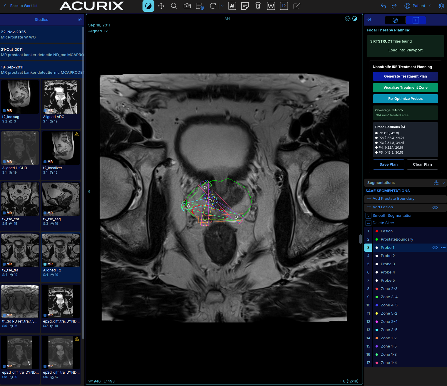
NanoKnife IRE (irreversible electroporation) treatment planning directly within the imaging viewer. Using the AI-generated prostate boundary and lesion segmentation as inputs, the system computes optimal probe placement to maximize lesion coverage with a safety margin.
Five probes are placed across four treatment phases, generating 10 treatment zones. Coverage fraction is computed in real time. Probes can be manually adjusted with zones recomputing automatically. The final plan is saved as a multi-slice DICOM RTSTRUCT for integration with treatment delivery systems.
Platform Capabilities
Cloud PACS
Integrated DCM4CHEE PACS with OHIF web viewer. Studies accessible from any browser without local DICOM software.
Multi-Site Ingestion
Connect existing PACS via DIMSE, SFTP, HTTP push, or C-MOVE pull. No changes to existing infrastructure required.
GPU Processing
AWS Batch with on-demand GPU instances. Models run on the latest NVIDIA hardware without any capital expenditure.
Multi-Tenant Isolation
Each organization gets cryptographically isolated data. Patient IDs are prefixed, S3 paths are segmented, and queries are filtered per tenant.
DICOM Standards
All results delivered as standard DICOM objects: RTSTRUCT for segmentations, DICOM SR for structured reports, standard series for aligned images.
Rapid Model Onboarding
Any containerized AI model can be deployed on the platform. Shared infrastructure for storage, compute, viewer integration, and HIPAA compliance.
HIPAA Compliance
Security and compliance are built into every layer, not bolted on afterward.
Encryption at Rest
AES-256 encryption for all stored data: S3, RDS, EFS. AWS KMS managed keys.
Encryption in Transit
TLS 1.2+ enforced across all connections. HSTS headers on all endpoints.
Audit Logging
Every PHI access event is logged to CloudWatch with 7-year retention. Full request tracing via AWS X-Ray.
The Acurix platform and the AI capabilities described on this page are under development and have not been cleared or approved by the U.S. Food and Drug Administration. They are not available for clinical use in the United States.